
Artificial Intelligence in Oncology: Clinical Applications, Challenges, and Opportunities
Am Soc Clin Oncol Educ Book. 2026 Jun;46(3):e520716.
OVERVIEW
Artificial intelligence (AI) is reshaping cancer research and clinical oncology by enabling large-scale analysis of complex biomedical data. Although early AI efforts focused on single-modality tasks such as imaging interpretation or pathology classification, contemporary oncology increasingly requires integrated reasoning across heterogeneous inputs, including imaging, digital pathology, and genomics. Multimodal artificial intelligence (MMAI) models represent a major evolution by fusing these data types into unified predictive systems capable of informing diagnosis, risk stratification, treatment selection, and disease monitoring. This manuscript reviews the state of AI in oncology and highlights emerging foundation models within the field. There will be a focus on histopathology, and how this can be incorporated into risk assessment models. There will also be examples of MMAI utility in radiology and spatial proteomics. Additionally, we will discuss ethical and legal implications of MMAI in modern oncology, and recommendations for responsible application within clinical oncology practice.
INTRODUCTION
Across medicine, artificial intelligence (AI) in medicine provides technical enhancement, which can contribute essential clinical impact. With defined intended use, rigorous external validation, calibrated outputs, workflow integration, and lifecycle monitoring, there are many opportunities for efficient translation within the clinical space.1 Oncology is uniquely positioned for AI because it generates highdimensional data and has mature clinical end points. However, these same characteristics can also create vulnerabilities: imaging protocols vary by site, slide preparation differs, molecular assays evolve, and standards of care change over time. Therefore, any AI system used in oncology must be designed not only to predict, but also to remain valid under real-world change.2,3
Multimodal AI (MMAI) can be integrated within oncology, as clinical decisions already synthesize multiple evidence streams within clinical outcomes, such as diagnostic imaging, pathology, genomics, and transcriptomics. Ideally, simultaneous MMAI models can combine clinical signals from a patient into a single quantitative framework (Fig 1— digital twin). This emerging concept supports dynamic clinical decisions, such as therapy escalation or deescalation, trial eligibility, and surveillance.4
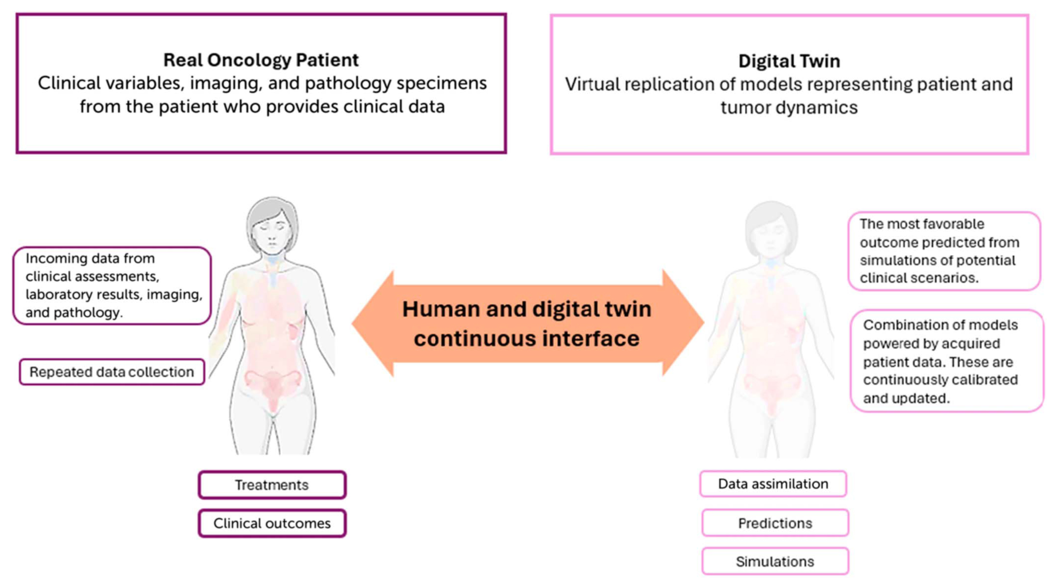
Fig 1. Patient with oncology care, alongside matching MMAI digital twin. This image shows the dual nature of the digital twin. Each patient provides continuous clinical data points into the digital twin, which uses MMAI to generate outcomes and guidelines for the real patient. MMAI, multimodal artificial intelligence.
In oncology, there have been several examples of MMAI models already deployed in clinic practice. For example, in non–small cell lung cancer, there have been developments of an AI-based model that applied serial computed tomography to generate radiomic biomarkers, to predict overall survival when cases were treated with immune checkpoint inhibitors.5 For breast cancer, the US Food and Drug Administration (FDA) recently approved Claire, an AI platform designed to assess breast cancer margins intra-operatively to support early surgical decisions.6 In pancreatic cancer, the Computational Histology AI platform, aimed to match the ideal first-line chemotherapy for advanced pancreatic ductal adenocarcinoma, with either fluoropyrimidine-based versus gemcitabine-based regimens, based on tumor histomorphology.7
For this manuscript, we will focus on how clinical data, histopathology, and imaging have been used to develop and validate MMAI models within oncology. However, in advance, we will discuss how MMAI in oncology can be used in a trustworthy manner through regulation and governance, using the ASCO policies as examples for guidelines.
TRUSTWORTHINESS IN ONCOLOGY AI: EVIDENCE, GOVERNANCE, AND REGULATION
Major AI-in-medicine reviews increasingly emphasize that the dominant risks in deployment are not only algorithmic, but sociotechnical: poorly defined intended use, hidden data set shift, workflow mismatch, and inadequate monitoring. Trustworthy oncology in conjunction with AI requires a lifecycle approach that spans development, validation, implementation, and continuous performance assurance.
PRACTICAL APPLICATIONS
- Our manuscript will provide knowledge and examples of multimodal artificial intelligence (MMAI) highlights in oncology, particularly in the field of histopathology, and how that can be incorporated into risk assessment models.
- Our manuscript will review current MMAI models in radiology and spatial proteomics.
- This paper discusses some ethical and legal implications of MMAI in modern oncology and provides recommendations for responsible application to clinical practice.
The Journal of Clinical Oncology defines AI in several broad groups for oncology: predictive AI, a system used to forecast an outcome based on data input placed into its system, and generative AI, which creates content based on learned patterns.8
With a dedicated taskforce, ASCO uses six principles when considering the utility of any proposed AI. These principles are transparency, informed stakeholders, fairness, accountability, oversight and privacy, and human-centered application. Through these principles, an AI tool can translate to assisting treatment decisions, not just predicting outcomes. Every AI system should be tied to a specific decision point and action. A prognostic score that does not change management has limited utility. In oncology, examples of actionability include selecting therapy intensification versus de-escalation, choosing imaging surveillance intervals, triggering confirmatory testing, and prioritizing trial enrollment.9
Importantly, oncology-associated AI systems must generalize across diverse populations, technical settings, and institutions. Therefore, equity should be a core requirement, as models that perform well overall but fail in specific underrepresented groups can worsen health care disparities. As a result, monitoring and adapting to accommodate all populations are essential. Oncology AI must be tracked for drift, including changes in case mix, imaging protocols, stains, assay versions, and treatment regimens. Governance should specify what can change, how performance will be revalidated, and how safety will be maintained. A practical governance toolkit includes automated performance dashboards, periodic chart review audits, drift detection triggers, and predetermined update plans.10,11
Regulatory expectations continue to evolve for AI-enabled software and medical devices. Oncology AI teams should be accountable for their designs through robust documentation, which should include intended use, training data provenance, validation of results, failure modes, human factors evaluation, and policy updates. When an AI component is integrated into a clinical pipeline, documentation should also specify how outputs will be used, what guardrails exist, and how errors will be handled.8
Therefore, to incorporate the above points, when considering the utilization of an MMAI, recommended key questions to contemplate include:
- Is this tool human-centered with a clear clinical function? Is it a predictive or a generative tool?
- Is the MMAI applicable to my core population?
- What are the guardrails used to account for accurate output and safeguard against errors?
- Is it equitable? Will it exacerbate existing gaps in oncology care?
- Is there transparency with how data are used between providers, patients, and other shareholders?
ROLE OF MMAI IN PATHOLOGY FOR DIAGNOSTICS
MMAI to Predict Clinical Outcomes From Genomic, Imaging, and Histopathology Data
Applications for Oncology
Although the number of FDA-approved AI tools for digital pathology is small, research in this area is wide-ranging and describes systems for diagnosis, grading and prognostication, treatment response prediction, and biomarker quantification.12–20 These tools aim to provide reproducible and often quantitative assessments that overcome interobserver variability and other limitations of visual interpretation. Advances in machine learning and computing in the 2010s produced powerful methods that can help us to identify highly predictive patterns in images. When applied to pathology, these methods can learn latent tissue patterns that predict characteristics, including somatic mutations and RNA expression. Although some of these patterns may be visible to pathologists, they are often not appreciated or reproducibly scorable.21,22
Cancer detection has been the primary focus of research and FDA-approved tools. The first FDA-cleared pathology image analysis system was approved in 1998 for cervical cancer screening.23 In 2021, Paige Prostate received FDA approval, demonstrating improvements in sensitivity and efficiency for detecting prostatic cancer in whole-slide images (WSIs) of biopsies, followed by Ibex Prostate Detect in 2025.
In 2025, ArteraAI received FDA approval for ArteraAI Prostate, which provides prognostication of clinical outcomes for patients diagnosed with localized prostate cancer. This tool infers prognosis from a WSI corresponding to the highest Gleason grade tissue block and the pathologist-determined grade. This system was validated in multiple phase 3 trials and shown to predict distant metastasis independent of other variables, including age, prostate-specific antigen, Gleason score, and T-stage.24 Prognostication is an active area of research, with emphasis on integrating data from multiple modalities.
Much of the work in predicting treatment response has focused on immunotherapy, given the relevance of the tumor microenvironment in determining response.25 Approaches to treatment response prediction include quantification of immunohistochemical staining of PD-L1, characterizing the abundance and distribution of tumor infiltrating lymphocytes, and learning latent patterns from images of hematoxylin and eosin (H&E)–stained slides.26 Immunotherapy response prediction is an ideal problem for AI, given the lack of predictive power of PD-L1 staining and the challenges in scoring including clonal heterogeneity, weak expression, and other factors.27
The slow adoption of AI tools in pathology reflects challenges in regulation, reimbursement, and providing requisite infrastructure. In the United States, the laboratory-developed test (LDT) framework permits diagnostic tests that are developed and validated in-house to be performed by individual laboratories.28 ArteraAI Prostate currently offers their test as an LDT through their clinical laboratory improvement amendments–certified laboratory, allowing laboratories that lack digital infrastructure to access their test. Developing strong evidence for test performance requires access to large and well-annotated observational studies and, ideally, prospective validation in clinical trials in the future.
Whole-Slide Imaging and Digital Pathology
Whole-slide imaging generates high-magnification images from glass slides, enabling digitization of pathology practices and the use of AI tools. Although whole-slide imaging has existed for decades, it was not until 2017 that the FDA approved the first digital pathology system for primary diagnosis of surgical pathology.29 Since then, subsequent studies have also demonstrated noninferior rates of major discordance for digital diagnosis relative to glass, resulting in the marketing of multiple FDA-approved digital pathology systems.30 The digital WSIs generated by these systems enable rapid remote consultation, and can improve costs, turnaround time, and quality by improving productivity, workload distribution, and reducing archive retrieval requests, lost slides, and misdiagnosis.31
European laboratories were early implementers of digital pathology, but in the United States, full digital implementation was slower and limited to several large academic centers. Digital implementation is costly, disruptive, and requires technical expertise. Archival storage and image management software programs are major cost drivers, given the size of WSIs, which are typically one or more gigabytes per slide. A fully digitized pathology laboratory can generate several million gigabytes of image data annually, requiring substantial on-premises or cloud storage, and information technology professionals to administer these systems. These issues may be particularly challenging for smaller laboratories that can benefit the most from digital pathology. More recently, reductions in the cost of implementation, proliferation of digital pathology systems, and a growing base of knowledge and experience have all contributed to accelerating adoption of digital pathology in the United States. Many of the benefits of digital pathology, such as consultation and AI, can be realized without substantial investments in storage for long-term retention (Fig 2).32
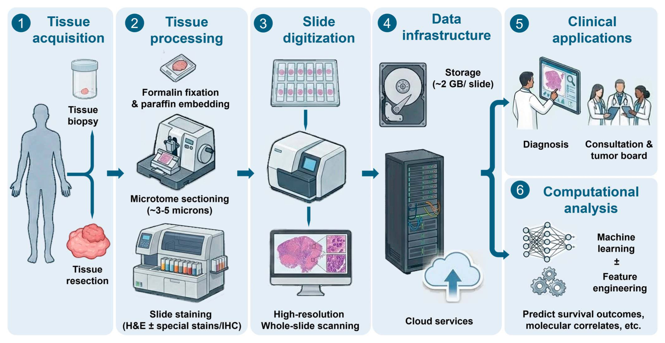
Fig 2. Overview of the digital pathology workflow, from tissue acquisition to clinical and computational applications. The process begins with tissue acquisition via biopsy or surgical resection, followed by standard processing steps such as formalin fixation, embedding in paraffin wax, microtome sectioning, and slide staining. Next, slide scanning generates high-magnification WSIs from the physical glass slides. Because individual WSIs are typically 1-3 gigabytes in size, fully digitized laboratories require substantial on-premises server infrastructure or cloud services to manage the massive amounts of data generated. Once digitized, these images seamlessly facilitate pathologist-driven clinical applications, such as primary diagnosis and rapid remote consultation. Furthermore, this digital infrastructure acts as the foundation for advanced computational analysis, including the extraction of quantitative patterns from WSIs to predict survival outcomes, treatment response, and molecular correlates. H&E, hematoxylin and eosin; IHC, immunohistochemistry; WSIs, whole-slide digital images.
Computational Pathology Paradigms
The dominant paradigm in developing computational pathology models is multi-instance learning (MIL), which uses patient-level labels to train AI models.33,34 The use of data as recorded in pathology reports avoids costly and subjective image annotation and enables access to data generated by pathology operations that exceed tens of thousands of slides. The basic elements of an MIL model are depicted in Figure 1. A whole-slide image is partitioned into tiles that correspond to the size of a typical high-power field. These tiles are processed by an encoder model that transforms the pixel values into a numerical representation of the entire tile. These representations are aggregated and further transformed by a model that adapts to identify which tiles are relevant to the patient-level label, and that weighs each tile appropriately. The encoder model is typically available in a pretrained form and is not adapted during this process, where the aggregator model is adapted.
The development of encoders is an active area of research that has yielded interesting results. Initially, encoders were borrowed from the general image analysis community, where they were trained to classify natural, nonmedical images. More recently, advances in a method called selfsupervised learning allow encoders to be trained using unlabeled data, leading to the development of pathology specific models that are trained on billions of high-power fields from millions of WSIs that represent diverse organs and conditions. Due to the breadth of their training data, these foundation encoders can be readily adapted to a wide variety of pathology applications, often with superior performance and less training data required.35,36
Another paradigm that predates MIL is feature engineering, where image segmentation is used to delineate tissue structures, and quantitative features are used to describe the morphologic properties of the tissue.37 Segmentation models are trained using annotations of tissue structures, which can be time-intensive and subjective, although the availability of publicly available annotation data has grown, and there are millions of annotations available for a wide variety of tissue contexts.38,39
These features are designed in a rational manner to capture prior knowledge about a disease or to encode a specific hypothesis and can provide transparency into the predictive mechanisms of AI models or explainability for specific predictions. Examples of features include the spatial distribution of tumor-infiltrating lymphocytes, morphology of tumor nuclei, properties of extracellular matrix, or topologic properties of ductal structures.40 Feature engineering is often used in prognostic or treatment predictive models, where there are strong priors and hypotheses about relevant patterns, and where interpretability and explainability beyond indications of salient regions is desired. A recent model for breast cancer prognosis developed a comprehensive feature set encompassing features of epithelial nuclei, glandular morphology, cancer-associated fibroblasts, extracellular matrix, tumor-infiltrating lymphocytes, and the interactions between these components, and novel associations with prognosis, immune response, and molecular subtypes of cancer-associated fibroblasts.38
Although feature engineering models are often considered to be less predictive than MIL, there is evidence that carefully constructed features can provide highly accurate predictions. Like MIL, feature engineering approaches have benefited significantly from recent advances in machine learning that improve image segmentation accuracy and that reduce training data requirements.41,42
Despite the success of these feature engineering strategies in extracting prognostic morphologic signatures, standard histopathology remains fundamentally constrained by its inability to identify specific protein targets and functional cellular states. As the field moves toward more personalized oncology, morphologic features alone offer an incomplete picture of the tumor microenvironment. Overcoming this limitation requires a modality that can explicitly measure high-dimensional protein expression while maintaining the structural tissue architecture learned through image analysis, a need directly addressed by the emerging integration of AI and spatial proteomics. As the following section details, this combined approach has become particularly transformative in breast cancer, where mapping the intricate spatial interplay within the microenvironment is essential for advancing targeted therapies.
Integrating Pathomics in MMAI-Driven Diagnostics in Breast Cancer
Spatial Proteomics and AI in Breast Cancer: From Tissue Architecture to Clinical Insight
In breast cancer, the clinical value of pathomics increasingly lies in its ability to capture tissue architecture and microenvironmental organization in ways that complement conventional molecular assays. This is important because the disease is defined not only by tumor-intrinsic features such as hormone receptor and human epidermal growth factor receptor 2 (HER2) status, but also by the spatial organization of tumor cells, immune infiltrates, and stroma. Traditional molecular assays average signals across many cells, limiting insight into how different cell populations are organized and interact within a tumor. Large-scale molecular profiling studies have demonstrated substantial biological heterogeneity in breast cancer, motivating techniques that preserve tissue context to better understand how these features are arranged within tumors.43 Spatial proteomics directly addresses this limitation by measuring protein expression in situ, which preserves tissue architecture and enables interrogation of cellular phenotypes, interactions, and higher-order tissue topology.44,45 and microenvironmental organization>When combined with AI, spatial proteomics provides a quantitative framework to extract clinically relevant patterns that may inform prognosis, therapeutic selection, and response assessment.46 Recent advances in AI, particularly foundation models and vision-language models, have accelerated the integration of spatial proteomics with histopathology and clinical data.35,47–49 These models offer a pathway from high-dimensional spatial measurements to interpretable outputs that align with clinical decision making. This section reviews spatial proteomics platforms, AI-driven analytic approaches, emerging multimodal foundation models, and their relevance to breast cancer care.
Spatial Proteomics Technologies Relevant to Breast Cancer
Spatial proteomics encompasses a range of imaging-based platforms capable of measuring dozens of protein markers while retaining spatial context. Such platforms include multiplex immunofluorescence, imaging mass cytometry, multiplexed ion beam imaging, GeoMx Digital Spatial Profiler from NanoString, MALDI immunohistochemistry (IHC), Expansion Proteomics (ProteomEx), and iterative imaging platforms such as CODEX.50–54 These platforms enable detailed characterization of immune cell subsets, tumor signaling states (eg, estrogen receptor [ER], HER2 pathway activation), and stromal components at single-cell resolution while also preserving spatial relationships that are critical for understanding tumor-immune interactions and microenvironmental organization.44,55
Clinical Applications and Discoveries
In breast cancer, spatial proteomics has revealed critical insights into breast cancer biology, including immune infiltration patterns, tumor-stroma interfaces, and cellular neighborhoods that are associated with prognosis and therapeutic response, particularly in triple-negative and HER2-positive disease.55–57 Multiregion spatial proteomic analyses demonstrated that proteomic heterogeneity increases with tumor progression independent of genomic heterogeneity and is closely linked to microenvironmental differences. Progression was characterized by increased macrophage and T-cell infiltration, upregulation of anti-inflammatory pathways such as kynurenine and prostaglandin signaling, and spatial features with prognostic significance. Increased lymphocyte infiltration was associated with improved survival in triple-negative and highly proliferative ER-positive breast cancers. Additionally, distinct immune-tumor neighborhood patterns were linked to favorable or poor outcomes.58,59
AI Integration With Spatial Proteomics
Machine Learning Approaches
Multiplex spatial images must be converted into structured data sets through cell segmentation, marker quantification, and quality control. Standardized pipelines enable reproducible processing of multiplex imaging data into single-cell spatial data sets, forming the foundation for downstream machine learning analyses.60 AI and machine learning are essential for analyzing the high-dimensional and spatially complex data sets generated by spatial proteomics platforms.44,61 Early approaches relied on cell-type frequencies or simple spatial metrics. More recent methods incorporate spatial proximity, clustering, and neighborhood structure to capture cell-cell interactions within intact tissue. Early machine learning approaches focused on extracting spatial features from in situ proteomic imaging to improve cancer survival modeling, typically relying on cell-type frequencies or simple spatial metrics. Although informative, these platforms provided limited insight into the organization of the tumor microenvironment. More recent methods, inspired by concepts from ecology and epidemiology, incorporate spatial proximity, clustering, and neighborhood structure to better capture cell-cell interactions within intact tissue.62
Deep learning approaches strategies, such as convolutional neural networks and graph-based spatial analyses, further enhance interpretation by supporting predictive modeling, biomarker discovery, and integration of spatial features with clinical variables.60,63 Advanced analytic frameworks incorporate spatial graph theory, topological data analysis, and agent-based modeling refine data integration and improve biological insight from spatial proteomics data sets, thereby laying the groundwork for more personalized therapeutic strategies.63
Foundation Models and Virtual Spatial Proteomics
Cost and scalability are the major limitations of spatial proteomics motivating the development of foundation models that learn generalizable tissue representations across data sets and modalities.43,44 These models capture reusable features that can be applied across multiple downstream tasks with minimal added supervision.
Large-scale pathology foundation models pretrained on millions of histology images generate transferable embeddings that can be aligned with spatial proteomics data, linking tissue morphology to cellular and molecular features. By providing a shared representational space, these models facilitate integration of routine histology, spatial proteomics, and clinical variables within a unified analytic framework. The emergence of vision-language foundation models represents an important advance for computational pathology and spatial biology. Models such as CONCH, MUSK, and mSTAR demonstrate how multimodal representations can improve performance across classification, molecular prediction, and outcome modeling tasks while enhancing interpretability through joint image-text-data reasoning.48,49,64
In breast cancer, these approaches offer a pathway to link routine histopathology with spatial proteomic features, enabling the identification of clinically relevant immune architectures, such as immune exclusion or organized tumor-immune niches, which may inform prognosis and therapeutic selection.
AI-Enabled Virtual Spatial Proteomics
An emerging extension is AI-enabled virtual spatial pro-teomics, in which spatial protein expression patterns are inferred directly from standard histopathology slides. HEX is a deep learning model trained on paired histology images and spatial proteomics data that can predict the expression of multiple immune and tumor biomarkers from routine H&E slides.65 Although initially validated in non–small cell lung cancer, this method illustrates a low-cost, scalable strategy to recover biologically meaningful spatial information and may enable broader access to spatial insights without requiring specialized experimental platforms (Fig 3).
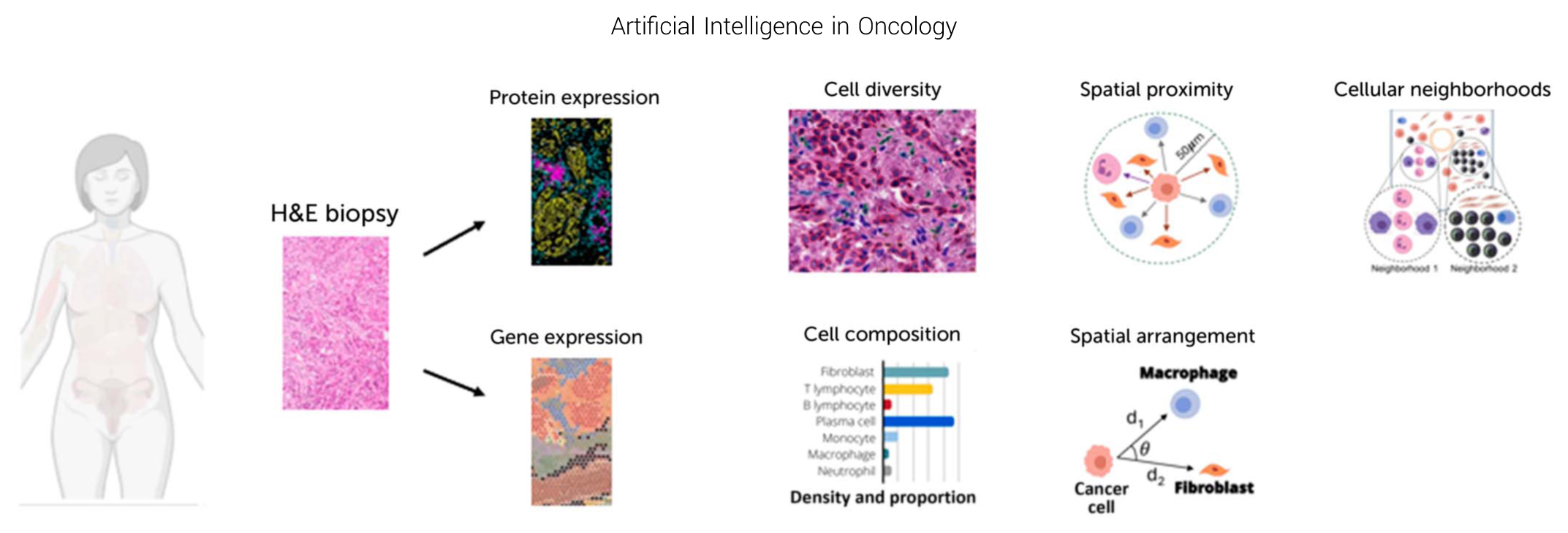
Fig 3. Spatial proteomics workflow for a patient with breast cancer. A diagnostic H&E biopsy from a patient with breast cancer serves as the entry point for spatially resolved molecular profiling. Spatial omics technologies generate complementary layers of information, including protein and gene expression, while preserving architecture. These molecular maps enable quantification of cell diversity and identification of spatial domains within the tumor microenvironment. Beyond measuring expression levels, spatial data support higher-order analyses of tissue organization (including cellular composition density and proportion of immune and stromal subsets), spatial proximity relationships within defined radii, cellular neighborhoods, and geometric arrangements among tumor, immune, and stromal cells. Together, these features provide a system-level view of tumor-microenvironment interactions that extend beyond histopathology and bulk molecular profiling. Image adapted from material provided Ryan Quinton MD, PhD, with permission. H&E, hematoxylin and eosin.
ctDNA Plus MMAI for Monitoring, Minimal Residual Disease, and Response-Adaptive Strategies
Complementing these tissue-based approaches, circulating tumor DNA (ctDNA) provides a minimally invasive and dynamic measure of disease burden and treatment response, making it an important component of multimodal AI strategies. ctDNA provides dynamic, biology-sensitive signals that complement imaging and pathology. It enables early detection of minimal residual disease, measurement of molecular response, and tracking of clonal evolution. Some current models have seen some difficulties in achieving primary end points, such as GRAIL in the National Health Service Galleri study, which failed to meet its primary end point of significantly reducing stage III to IV cancer diagnoses.66,67 A possible next step could be multimodal integration: combining ctDNA trajectories with imaging features and pathology-derived representations to improve prediction of recurrence, resistance, and outcomes, which can guide response-adaptive care strategies.
In multicancer early detection, CANSCAN is a test that uses whole-genome sequencing and AI to analyze cell-free DNA in an effort to facilitate earlier diagnoses and more accurate tissue origins. From the DECIPHER-Omnia study, CANSCAN was developed and screened for both independent and asymptomatic cohorts. The DECIPHER-Omnia study was undertaken in three phases: model development (in an internal validation cohort), independent validation (in a separate cohort), and validation in an asymptomatic screening population.68
In the first phase, 3,076 patients provided blood samples with cancer (13 types) and 3,477 patient samples were collected for age-matched healthy controls (divided into a training set [n = 4,807] and an internal validation set [n = 1,746]).The second phase of 1,465 participants (732 with and 733 without cancer) involved an independent validation with comparable baseline demographics across cohorts. In independent validation, CANSCAN achieved 87.4% overall sensitivity and 97.8% specificity across 13 cancer types, alongside 91.7% tissue-of-origin prediction accuracy. For early-stage cancers, at stage I, there was a 79.3% sensitivity, and at stage II, there was an 86.9% sensitivity. For the screening cohort (3,724 asymptomatic individuals age between 45 and 75 years), sensitivity was 53.3% and specificity was 98.1%. The third phase is a planned prospective cohort study, with estimation of enrolling 15,000 asymptomatic individuals.68
For ovarian cancer, DNA evaluation of fragments for early interception (DELFI) applies to the patterns in circulating DNA fragments found in liquid biopsy analysis to integrate the fragmentome data with plasma levels of two known biomarkers of ovarian cancer: the proteins CA125 and HE4. This emerging diagnostic model aims to differentiate between benign and malignant ovarian tumors, with another model to screen asymptomatic patients. With a small sample size of 134 women with ovarian cancer, 204 women without cancer, and 203 women with benign adnexal masses, DELFI demonstrated specificity of over 99%. The screening model identified 69%, 76%, 85%, and 100% of ovarian cancer cases staged I to IV, respectively, compared with an analysis of CA125 levels alone, which identified 40%, 66%, 62%, and 100% of cases, respectively.69
ROLE OF MMAI IN PATHOLOGY FOR CLINICAL RISK ASSESSMENT AND APPLICATIONS
Using Clinical and Biospecimens Data to Validate AI-Driven Diagnostics and Risk Assessment
AI risk models become clinically meaningful only when they demonstrate analytic validity, clinical validity, and clinical utility. Cooperative group trials and well-annotated biobanks are uniquely powerful for oncology AI validation because treatments are protocol-defined, end points are prespecified and adjudicated, and follow-up is systematic. These characteristics enable robust evaluation of prognostic performance, predictive performance for treatment benefit, calibration, and subgroup equity.
Prognostic Factors for Recurrence After Local Therapy Guide Adjuvant Systemic Therapy
ER-positive, HER2-negative, node-negative breast cancer accounts for about one half of all breast cancers in the United States. Although it is associated with generally favorable prognostics and low recurrence risk, it accounts for about 60% of all breast cancer deaths due to its high prevalence and persistent recurrence risk beyond 5 years.70 Distant recurrences may occur over 30 years after potentially curative surgery, despite adjuvant systemic therapy. Although adjuvant systemic chemotherapy and endocrine therapy reduce recurrence risk, with chemotherapy reducing recurrences largely within the first 5 years, more than half of distant recurrences occur more than 5 years after initial surgery, often after completion of a 5-year or longer course of adjuvant antiestrogen endocrine therapy.70,71
Gene Expression Assay as a Prognostic Tool in Hormone Receptor-Positive, HER2-Negative Breast Cancer
The Oncotype 21-gene recurrence score (RS) provides prognostic information for distant recurrence and predictive information for chemotherapy benefit in ER-positive, HER2-negative early breast cancer (Table 1).76–81
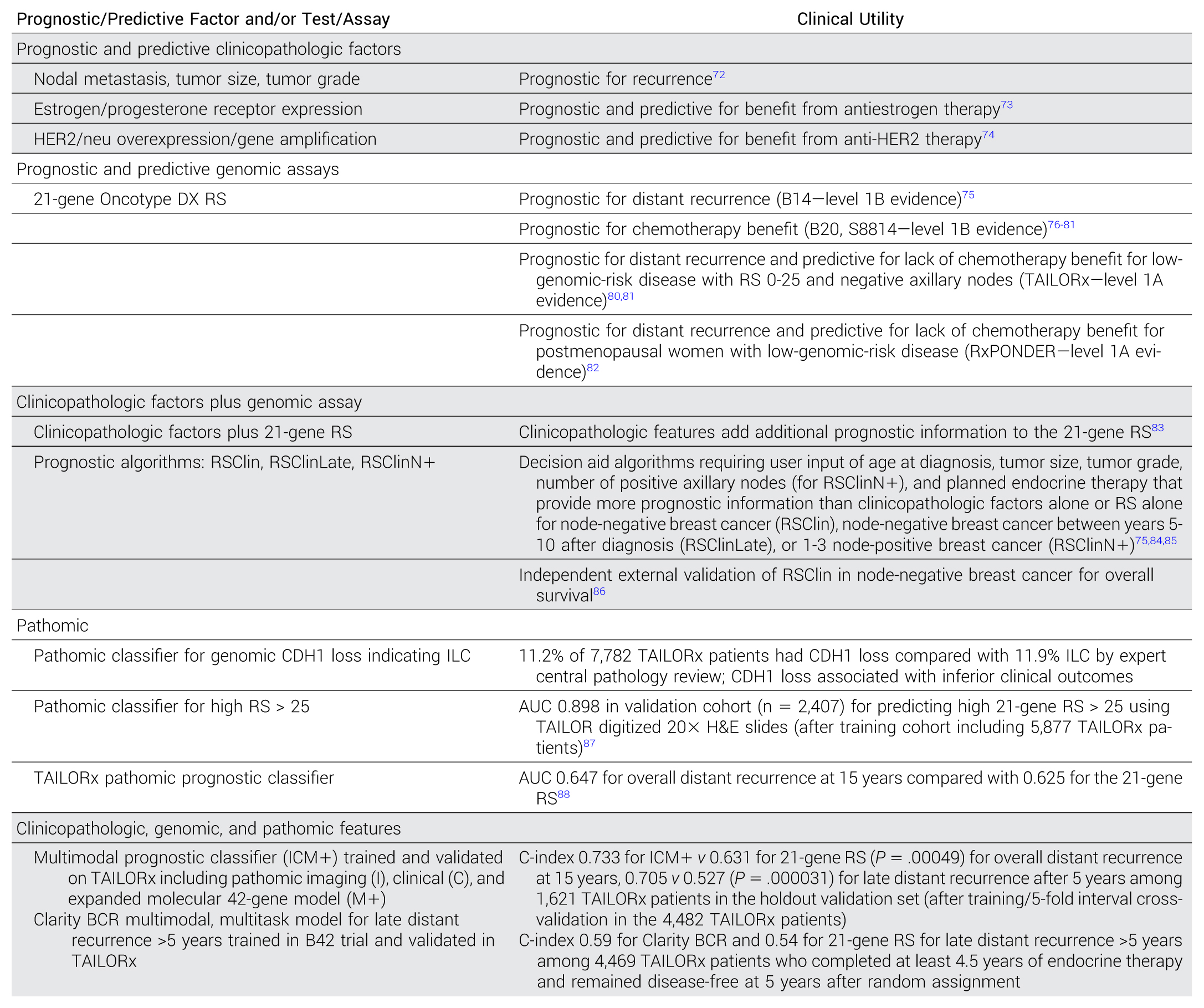
Table 1. Prognostic and Predictive Factors for Localized Breast Cancer in Clinical Trial Cohorts, Including Cohorts Evaluating the 21-Gene RS and Pathomic Models
Abbreviations: BCR, breast cancer recurrence; H&E, hematoxylin and eosin; HER2, human epidermal growth factor receptor 2; ICM, pathomic imaging (I), clinical (C), and expanded molecular 42-gene model (M); ILC, invasive lobular carcinoma; RS, recurrence score; RxPONDER, Rx for Positive Node, Endocrine Responsive Breast Cancer; TAILORx, Trial Assigning Individualized Options for Treatment.
The Trial Assigning Individualized Options for Treatment (TAILORx) established the clinical utility of the 21-gene RS for providing prognostic information for distant recurrence and guiding chemotherapy use in ER-positive, HER2-negative, axillary node–negative breast cancer, and has been recommended for this purpose by multiple practice guidelines dating back to 2019. Clinicopathologic features add additional prognostic information to the 21-gene RS.80,81,83,87,89–91 The Clinical Trial Rx for Positive Node, Endocrine Responsive Breast Cancer demonstrated that the RS may also be used to guide chemotherapy in postmenopausal women with ER-positive, HER2-negative breast cancer with 1-3 positive axillary nodes.82 Decision aid algorithms integrating prognostic information derived from clinicopathologic factors and the RS provide more prognostic information that either alone for nodenegative breast cancer (RSClin), node-negative breast cancer between years 5-10 after diagnosis (RSClinLate), and 1-3 node-positive breast cancer (RSClinN+). Independent external validation of RSClin in node-negative breast cancer for chemotherapy benefit has also been shown.75,84–86
Development and/or Validation of Multimodal Prognostic Models in TAILORx
Pathomic classifiers in breast cancer assess a range of pathologic features extracted from digitized H&E slides, either alone or integrated with gene expression–based and/or clinical data, to enhance prediction of recurrence risk. Several pathology foundation models have been developed to improve interpretation of predictive biomarkers. Although their performance has been variable, their accuracy has improved.
A secondary objective of TAILORx was to develop a biospecimen repository for using clinical data and formalin-fixed, paraffin-embedded archival primary tumor samples derived from trial participants. As summarized in Table 1, TAILORx clinical trial biospecimens and data were leveraged to develop and prospectively validate multimodal models integrating clinical, molecular, and pathomic imaging features to provide stronger discriminatory prognostic information for distant recurrence that any of those features used individually.
Other Pathomic Classifiers
Other pathomic classifiers may serve as a surrogate for prognostic and/or predictive biomarkers or add prognostic information when integrated with gene expression assays or clinicopathologic factors, as summarized in Table 2 and described below.
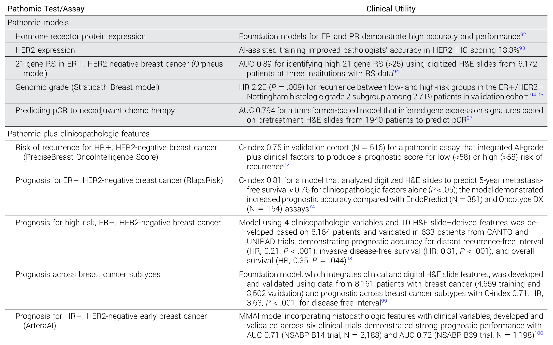
Table 2. Other Pathomic Classifiers
Abbreviations: ER, estrogen receptor; H&E, hematoxylin and eosin; HER2, human epidermal growth factor receptor 2; HR, hazard ratio; IHC, immunohistochemistry; MMAI, multimodal artificial intelligence; pCR, pathologic complete response; PR, progesterone receptor; RS, recurrence score.
Pathomic Surrogate for Prognostic and Predictive Biomarkers
In a systematic comparison of public pathology foundation models, the AUC for ER expression ranged from 0.95 to 0.97, and for progesterone receptor expression from 0.89 to 0.9375.92 A training platform for AI-supported digital HER2 IHC assessment of breast cancer samples improved pathologists’ accuracy in HER2 IHC scoring by 13.3%, and decreased misclassification of HER2-ultralow cases as HER2 null.93
Regarding histologic grade, the Stratipath Breast model uses digital H&E-stained WSIs to improve risk stratification of patients with Nottingham histologic grade 2 breast cancer. In a validation set of over 2,000 patients, among those with grade 2, ER-positive, HER2-negative breast cancer, the hazard ratio (HR) for recurrence was 2.2 (P = .009) between low- and high-risk groups stratified by the model, suggesting its ability to improve risk stratification.95,96
Regarding gene expression biomarkers, the Orpheus model infers the 21-gene RS from digitized H&E slides. In a validation set of 6,172 cases, the model identified cases of high RS (> 25) with an AUC of 0.89, compared with a leading clinicopathologic nomogram with AUC 0.73. Among patients with RS ≤ 25, the model predicted the risk of distant recurrence more accurately than RS alone, with AUC of 0.75 versus 0.49, respectively.94 Similarly, Howard et al applied transformer-based models to pretreatment H&E biopsy slides from 1,940 patients with breast cancer treated with neoadjuvant chemotherapy to infer gene expression signatures predictive of pathologic complete response (pCR). The most predictive model was derived from I-SPY2 trial participants and validated in four external cohorts. Among those with HER2-negative breast cancer, the transformer-based model predicted pCR with an AUC of 0.794, outperforming models based on clinical features alone (AUC 0.704, P = .001).97
Pathomics Plus Clinicopathologic Factors
Several models integrating prognostic information from pathomics plus clinicopathologic factors have been developed and validated.
The PreciseBreast OncoIntelligence Score analyzes digitized H&E slides and clinical characteristics to enhance breast cancer grading and prognostication; patients are assigned a risk score from 0 to 100 and stratified by low (<58) or high (>58) risk of recurrence. The model trained in 1,559 patients had a C-index of 0.78, which was superior to clinical (C-index 0.71) and imaging (C-index 0.72) feature models. In the validation cohort of 516 patients, the C-index for the model (0.75) remained superior to clinical (C-index 0.71) and imaging (C-index 0.67) feature models.72
The RlapsRisk BC model evaluates digitized H&E slides to predict 5-year metastasis-free survival in ER-positive, HER2-negative early breast cancer. The model had higher prognostic accuracy than clinicopathologic variables alone (C-index 0.81 v 0.76, P < .05).73 The model also had improved performance for predicting 5-year distant recurrence-free survival compared with the EndoPredict (AUC 0.73 v 0.57) and the 21-gene RS (AUC 0.78 v 0.58).74
Bidard et al demonstrated the clinical utility of a model integrating four clinicopathologic variables with 10 features from digitized H&E slides, which capture tumor architecture, microenvironment, and proliferation, to refine risk for patients with high-risk, HR-positive, HER2-negative early breast cancer. The model was developed based on 6,164 patients and classifies patients as low-risk or not low-risk of recurrence. Validation in 633 high-risk patients on the prospective trials CANTO and UNIRAD demonstrated that 95.4% of low-risk patients remained free of distant recurrence and death at 9 years compared with 76.8% for the not low-risk group. The assay predicted distant recurrence-free interval (HR, 0.21 [95% CI, 0.09 to 0.52]; P < .001), invasive disease-free survival (HR, 0.31 [95% CI, 0.16 to 0.60]; P < .001), and overall survival (HR, 0.31 [95% CI, 0.16 to 0.60]; P < .001).98
Another foundation model based on both digital H&E slides and clinical features has been shown to be prognostic across breast cancer types. The test was developed and evaluated using data from 8,161 patients with breast cancer across 15 cohorts. Of these, 3,502 patients from five cohorts were used for evaluation, while the remaining were used for training. The test accurately predicted disease-free interval, with a C-index of 0.71 (95% CI, 0.68 to 0.75) and HR of 3.63 ([95% CI, 3.02 to 4.37]; P < .001) for high- versus low-risk groups. In a direct comparison (N = 858), the AI test was more accurate than the 21-gene RS (C-index, 0.67 v 0.61). Furthermore, the AI test added independent prognostic information to the 21-gene RS in a multivariate analysis (HR, 3.11 [95% CI, 1.91 to 5.09]; P < .001). The model was also prognostic for triple-negative breast cancer (C-index 0.71, HR, 3.81 [95% CI, 2.35 to 6.17]; P = .02).99
Artera MMAI Platform Overview
MMAI models developed by Artera integrate digital pathology, clinical variables, and longitudinal outcomes to generate prognostic and predictive tools applicable to real-world treatment decisions. These models represent a practical example of multimodal AI translation, combining histopathologic representations with structured clinical data to support risk stratification and therapeutic selection. Importantly, their development and validation have leveraged large phase III clinical trial data sets with standardized end points and long-term follow-up, making these models particularly relevant to oncology practice.24
Application for Prostate Cancer
ArteraAI Prostate Test is a National Comprehensive Cancer Network (NCCN) guideline-recommended biomarker test for localized prostate cancer. It uses a MMAI algorithm, trained on digitized pathology slides and clinical data derived from multiple phase III randomized clinical trials, which evaluated radiotherapy with or without androgen-deprivation therapy. This provided individualized 10-year risk predictions for distant metastasis and prostate cancer–specific mortality in efforts to guide treatment decisions. This algorithm was trained on five phase III cohorts (NRG/RTOG 9202, 9408, 9413, 9910, and 0126), which combined 16,000 total biopsies from original diagnostic and subsequent treatment outcomes.12,88
ArteraAI Prostate Test requires from each patient a H&E-stained biopsy slide and the associated clinical variables. Through this test, the algorithm can provide insights into active surveillance eligibility for patients classified as very low, low, or favorable-intermediate risk by NCCN guidelines. For example, in 292 patients, MMAI scores predicted those who with higher MMAI scores would have adverse pathologic characteristics. These patients started on active surveillance but later required radical prostatectomy.88,101
Also, the test helps recognize which intermediate-risk patients (according to NCCN) would benefit from adding short-term androgen-deprivation therapy to radiation. Specifically, the predicative model to guide the benefit of short-term Androgen Deprivation Therapy (ADT) was trained on 3,977 patients. It showed that the 15-year metastasis risk was 9.8% for patients treated with radiotherapy alone versus 5.9% with ADT plus radiotherapy in the entire cohort. For the biomarker-positive patients, this model found a substantial benefit from ADT, while biomarker-negative patients saw minor difference. This proved the ability to identify subsets deriving intensified androgen-deprivation therapy in combination with radiotherapy.
ArteraAI Prostate Test also helped select patients classified as NCCN high-risk localized disease, who would gain the most from hormone therapy intensification such as combining abiraterone with long-term androgen-deprivation therapy. With an independent cohort and the STAMPEDE cohort (randomized trial that demonstrated that the addition of a second-generation hormone therapy to the standard-of-care treatment arm improved overall and metastasis-free survival in patients with high-risk nonmetastatic prostate cancer), ArteraAI showed that the MMAI risk score predicted patient outcomes. MMAI risk scores that were higher were associated with patients who benefited from combining abiraterone with standard-of-care hormonal therapy.102–105
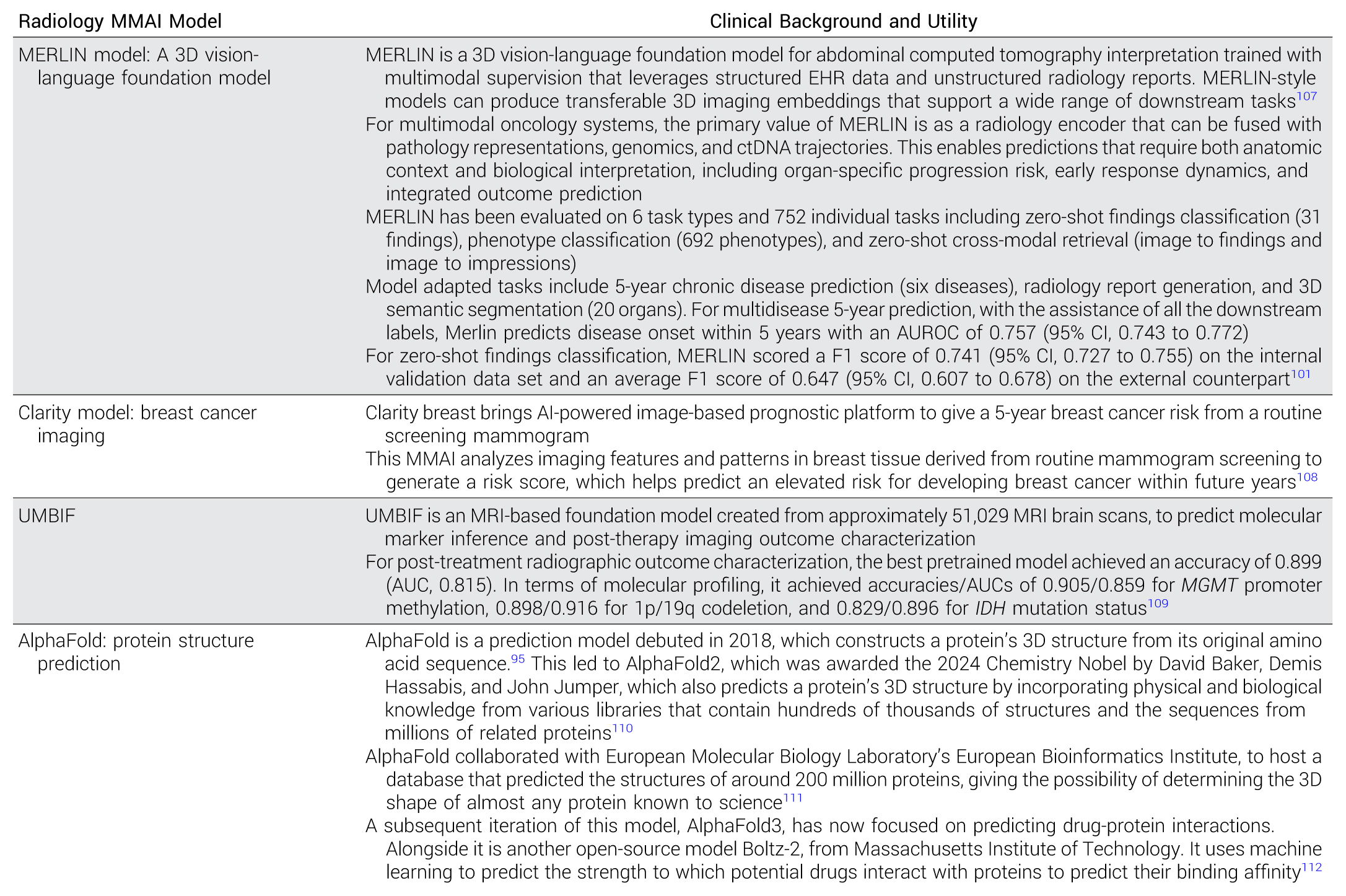
Table 3.Sample MMAI Models in Radiology and Protein Modeling
Abbreviations: AI, artificial intelligence; AUROC, area under the receiver operating characteristic; ctDNA, circulating tumor DNA; EHR, electronic health record; MMAI, multimodal artificial intelligence; MRI, magnetic resonance imaging; UMBIF, Unified Multimodal Brain Imaging Foundation.
Application in Breast Cancer: NSABP B-20 and Related Cohorts
In hormone receptor–positive, node-negative, early-stage breast cancer, decisions regarding chemotherapy use remain challenging, particularly in postmenopausal populations. The ArteraAI MMAI pathomic model was developed using tumor specimen derived from 8,616 patients enrolled in four trials (ADAPT, PlanB, NSABP B-34, and ABCSG 6). The model produces a continuous MMAI raw score, which ranges from 0 to 100, and was validated in two clinical trial cohorts with ER-positive breast cancer enrolled in NSABP B39 (n = 1,198) and the tamoxifen arm of NSABP B14 (n = 2,188); MMAI demonstrated better prognostic performance compared with clinicopathologic features alone in B14. The MMAI model has been externally validated using risk-stratified groups of low, intermediate, and high risk of distant recurrence, and it was found to be prognostic of 10-year recurrence risk in 1,763 patients from the NSABP B-20 trial, who were randomly assigned to receive adjuvant tamoxifen or chemotherapy plus tamoxifen.106
These findings illustrate how MMAI approaches may complement or refine existing clinicopathologic and genomic risk assessment strategies by incorporating morphologic and microenvironmental features captured in routine histology. Ongoing efforts aim to further validate MMAI performance across additional data sets and integrate such tools into prospective decision frameworks.
ROLE OF MMAI IN DIAGNOSTIC RADIOLOGY AND PROTEIN MODELING
Radiology is central to oncology for diagnosis, staging, response assessment, and surveillance. Although still new, MMAI has progressed from narrow detectors to generalizable segmentation and foundation-model representations. In practical oncology workflows, radiomics can provide an MMAI-centric combination of volumetric assessment, longitudinal tracking, and more consistent response measurement. Emerging in the protein modeling space, MMAI has allowed for the recreation of natural protein structure based on the amino acid building blocks and the archived data available on previous proteins investigated. The table below shows several MMAI models in radiology and protein modeling (Table 3).
CONCLUSION
In conclusion, MMAI can become an integral part of oncology intake processes and decisions. However, AI systems should be developed responsibly to ensure that they can be patient-centered, clinically useful, reliable, and equitable. Although we have shared some leading examples on how MMAI models have been applied in oncology, particularly in the fields of clinical data, pathology, and imaging, the list continues to be extensive, as MMAI evolves in the oncology-associated clinical space.
REFERENCES
Obermeyer Z, Emanuel EJ: Predicting the future—Big data, machine learning, and clinical medicine. N Engl J Med 375:1216-1219, 2016 | |
Lotter W, Hassett MJ, Schultz N, et al: Artificial intelligence in oncology: Current landscape, challenges, and future directions. Cancer Discov 14:711-726, 2024 | |
Luchini C, Pea A, Scarpa A: Artificial intelligence in oncology: Current applications and future perspectives. Br J Cancer 126:4-9, 2022 | |
Asghar US, Chung C: Application of digital twins for personalized oncology. Nat Rev Cancer 25:823-825, 2025 | |
Sako C, Kurland BF, Schmidt TG, et al: Deep-learning serial CT prediction of survival in immunotherapy-treated non-small cell lung cancer. JAMA Netw Open 9:e2555759, 2026 | |
ASCO AI Staff: First AI-Enabled Imaging Device for Breast Cancer Surgery Granted FDA Premarket Approval. https://ascoai.org/articles/2026/03/first-ai-enabled-imaging-device-for-breast-cancer-surgery-granted-fda-premarket-approval | |
Hendifar AE, Krishna V, Krishna V, et al: Development and validation of a computational histology artificial intelligence-powered predictive biomarker for selection of chemotherapy in advanced pancreatic cancer. J Clin Oncol 10.1200/JCO-25-02199 [epub ahead of print on February 2026] | |
American Society of Clinical Oncology (ASCO): Principles for the Responsible Use of Artificial Intelligence in. AI and Oncology, 2025. https://cdn.bfldr.com/KOIHB2Q3/as/g5jsnp7g2b6m28j67j97smff/2025-ASCO-AI-Principles | |
Parikh RB, Iasonos A, Ko A, et al: Bringing trustworthy artificial intelligence to the clinical forefront at JCO: A guide for studies testing artificial intelligence models. J Clin Oncol 43:3434-3438, 2025 | |
Obermeyer Z, Powers B, Vogeli C, et al: Dissecting racial bias in an algorithm used to manage the health of populations. Science 366:447-453, 2019 | |
Parikh RB, Teeple S, Navathe AS: Addressing bias in artificial intelligence in health care. JAMA 322:2377-2378, 2019 | |
Esteva A, Feng J, van der Wal D, et al: Prostate cancer therapy personalization via multi-modal deep learning on randomized phase III clinical trials. NPJ Digit Med 5:71, 2022 | |
Raciti P, Sue J, Retamero JA, et al: Clinical validation of artificial intelligence-augmented pathology diagnosis demonstrates significant gains in diagnostic accuracy in prostate cancer detection. Arch Pathol Lab Med 147:1178-1185, 2023 | |
Pantanowitz L, Quiroga-Garza GM, Bien L, et al: An artificial intelligence algorithm for prostate cancer diagnosis in whole slide images of core needle biopsies: A blinded clinical validation and deployment study. Lancet Digit Health 2:e407-e416, 2020 | |
Cantley RL, Jing X, Smola B, et al: Validation of AI-assisted ThinPrep® Pap test screening using the Genius™ Digital Diagnostics System. J Pathol Inform 15:100391, 2024 | |
Lu MY, Chen TY, Williamson DFK, et al: AI-based pathology predicts origins for cancers of unknown primary. Nature 594:106-110, 2021 | |
Sandbank J, Bataillon G, Nudelman A, et al: Validation and real-world clinical application of an artificial intelligence algorithm for breast cancer detection in biopsies. NPJ Breast Cancer 8:129, 2022 | |
Shen J, Choi YL, Lee T, et al: Inflamed immune phenotype predicts favorable clinical outcomes of immune checkpoint inhibitor therapy across multiple cancer types. J Immunother Cancer 12: e008339, 2024 | |
Cortellini A, Zampacorta C, De Tursi M, et al: A preliminary study on the diagnostic performance of the uPath PD-L1 (SP263) artificial intelligence (AI) algorithm in patients with NSCLC treated with PD-1/PD-L1 checkpoint blockade. Pathologica 116:222-231, 2024 | |
Krishnamurthy S, Schnitt SJ, Vincent-Salomon A, et al: Fully automated artificial intelligence solution for human epidermal growth factor receptor 2 immunohistochemistry scoring in breast cancer: A multireader study. JCO Precis Oncol 10.1200/PO.24.00353 | |
Echle A, Grabsch HI, Quirke P, et al: Clinical-grade detection of microsatellite instability in colorectal tumors by deep learning. Gastroenterology 159:1406-1416.e11, 2020 | |
Hallinan C, Lucas CHG, Fan J: Impact of data quality on deep learning prediction of spatial transcriptomics from histology images. bioRxiv 10.1101/2025.09.04.674228 | |
Bengtsson E, Malm P: Screening for cervical cancer using automated analysis of PAP-smears. Comput Math Methods Med 2014:842037, 2014 | |
Spratt DE, Liu VYT, Jia AY, et al: Meta-analysis of individual patient-level data for a multimodal artificial intelligence biomarker in high-risk prostate cancer: Results from six NRG/RTOG phase 3 randomized trials. Eur Urol 86:369-371, 2024 | |
Zhang J, Choi H, Kim Y, et al: Artificial intelligence-based digital pathology using H&E-stained whole slide images in immuno-oncology: From immune biomarker detection to immunotherapy response prediction. J Immunother Cancer 13:e011346, 2025 | |
Schuiveling M, van Duin IAJ, Ter Maat LS, et al: Artificial intelligence-detected tumor-infiltrating lymphocytes and outcomes in Anti-PD-1-Based treated melanoma. JAMA Oncol 11:1470-1478, 2025 | |
Liu Y, Chen W, Ruan R, et al: Deep learning based digital pathology for predicting treatment response to first-line PD-1 blockade in advanced gastric cancer. J Transl Med 22:438, 2024 | |
Aggarwal A, Bharadwaj S, Corredor G, et al: Artificial intelligence in digital pathology—Time for a reality check. Nat Rev Clin Oncol 22:283-291, 2025 | |
Mukhopadhyay S, Feldman MD, Abels E, et al: Whole slide imaging versus microscopy for primary diagnosis in surgical pathology: A multicenter blinded randomized noninferiority study of 1992 shenCases (Pivotal Study). Am J Surg Pathol 42:39-52, 2018 | |
Borowsky AD, Miller DV, Bauer TW, et al: A multicenter study to evaluate diagnostic accuracy by pathologists using the Aperio GT 450 DX in local and remote viewing stations. Arch Pathol Lab Med 149:859-867, 2025 | |
Hanna MG, Reuter VE, Samboy J, et al: Implementation of digital pathology offers clinical and operational increase in efficiency and cost savings. Arch Pathol Lab Med 143:1545-1555, 2019 | |
Zarella MD, McClintock DS: Don’T Be Afraid of Storage Costs. Digital Pathology Association, 2023. https://digitalpathologyassociation.org/blog/dont-be-afraid-of-storage-costs | |
Campanella G, Hanna MG, Geneslaw L, et al: Clinical-grade computational pathology using weakly supervised deep learning on whole slide images. Nat Med 25:1301-1309, 2019 | |
Lu MY, Williamson DFK, Chen TY, et al: Data-efficient and weakly supervised computational pathology on whole-slide images. Nat Biomed Eng 5:555-570, 2021 | |
Chen RJ, Ding T, Lu MY, et al: Towards a general-purpose foundation model for computational pathology. Nat Med 30:850-862, 2024 | |
Xu H, Usuyama N, Bagga J, et al: A whole-slide foundation model for digital pathology from real-world data. Nature 630:181-188, 2024 | |
Bera K, Schalper KA, Rimm DL, et al: Artificial intelligence in digital pathology—New tools for diagnosis and precision oncology. Nat Rev Clin Oncol 16:703-715, 2019 | |
Amgad M, Elfandy H, Hussein H, et al: Structured crowdsourcing enables convolutional segmentation of histology images. Bioinformatics 35:3461-3467, 2019 | |
Liu S, Amgad M, More D, et al: A panoptic segmentation dataset and deep-learning approach for explainable scoring of tumor-infiltrating lymphocytes. NPJ Breast Cancer 10:52, 2024 | |
Fassler DJ, Torre-Healy LA, Gupta R, et al: Spatial characterization of tumor-infiltrating lymphocytes and breast cancer progression. Cancers (Basel) 14:2148, 2022 | |
Amgad M, Hodge JM, Elsebaie MAT, et al: A population-level digital histologic biomarker for enhanced prognosis of invasive breast cancer. Nat Med 30:85-97, 2024 | |
Leo P, Janowczyk A, Elliott R, et al: Computer extracted gland features from H&E predicts prostate cancer recurrence comparably to a genomic companion diagnostic test: A large multi-site study. NPJ Precis Oncol 5:35, 2021 | |
Curtis C, Shah SP, Chin SF, et al: The genomic and transcriptomic architecture of 2,000 breast tumours reveals novel subgroups. Nature 486:346-352, 2012 | |
Keren L, Bosse M, Marquez D, et al: A structured tumor-immune microenvironment in triple-negative breast cancer revealed by multiplexed ion beam imaging. Cell 174:1373-1387.e19, 2018 | |
Walker C, Angelo M: Toward clinical applications of spatial-omics in cancer research. Nat Cancer 5:1771-1773, 2024 | |
McKenzie M, Irac SE, Chen Z, et al: Integrative spatial omics and artificial intelligence: Transforming cancer research with omics data and AI. Semin Cancer Biol 119:65-82, 2026 | |
Noorbakhsh J, Foroughi Pour A, Chuang J: Emerging AI approaches for cancer spatial omics. GigaScience 14:giaf128, 2025 | |
Lu MY, Chen B, Williamson DFK, et al: A visual-language foundation model for computational pathology. Nat Med 30:863-874, 2024 | |
Xiang J, Wang X, Zhang X, et al: A vision-language foundation model for precision oncology. Nature 638:769-778, 2025 | |
Marx V: Method of the year: Spatially resolved transcriptomics. Nat Methods 18:9-14, 2021 | |
Vandereyken K, Sifrim A, Thienpont B, et al: Methods and applications for single-cell and spatial multi-omics. Nat Rev Genet 24:494-515, 2023 | |
Bergholtz H, Carter JM, Cesano A, et al: Best practices for spatial profiling for breast cancer research with the GeoMx Digital Spatial Profiler. Cancers 13:4456, 2021 | |
Li L, Sun C, Sun Y, et al: Spatially resolved proteomics via tissue expansion. Nat Commun 13:7242, 2022 | |
Claes BSR, Krestensen KK, Yagnik G, et al: MALDI-IHC-guided in-depth spatial proteomics: Targeted and untargeted MSI combined. Anal Chem 95:2329-2338, 2023 | |
Jackson HW, Fischer JR, Zanotelli VRT, et al: The single-cell pathology landscape of breast cancer. Nature 578615-620, 2020 | |
Houlahan KE, Mangiante L, Sotomayor-Vivas C, et al: Complex rearrangements fuel ER+ and HER2+ breast tumours. Nature 638:510-518, 2025 | |
Greenwald NF, Nederlof I, Sowers C, et al: Temporal and spatial composition of the tumor microenvironment predicts response to immune checkpoint inhibition. bioRxiv 10.1101/2025.01.26.634557 | |
Mardamshina M, Karagach S, Mohan V, et al: Integrated spatial proteomic analysis of breast cancer heterogeneity unravels cancer cell phenotypic plasticity. Nat Commun 16:10482, 2025 | |
Eng JR, Bucher E, Hu Z, et al: Highly multiplexed imaging reveals prognostic immune and stromal spatial biomarkers in breast cancer. JCI Insight 10:e176749, 2025 | |
Schapiro D, Jackson HW, Raghuraman S, et al: histoCAT: Analysis of cell phenotypes and interactions in multiplex image cytometry data. Nat Methods 14:873-876, 2017 | |
Mou M, Pan Z, Lu M, et al: Application of machine learning in spatial proteomics. J Chem Inf Model 62:5875-5895, 2022 | |
Dayao MT, Trevino A, Kim H, et al: Deriving spatial features from in situ proteomics imaging to enhance cancer survival analysis. Bioinformatics 39:i140-i148, 2023 (39 Suppl 1) | |
Rajdeo P, Aronow B, Surya Prasath VB: Deep learning-based multimodal spatial transcriptomics analysis for cancer. Adv Cancer Res 163:1-38, 2024 | |
Xu Y, Wang Y, Zhou F, et al: A multimodal knowledge-enhanced whole-slide pathology foundation model. Nat Commun 16:11406, 2025 | |
Li Z, Li Y, Xiang J, et al: AI-enabled virtual spatial proteomics from histopathology for interpretable biomarker discovery in lung cancer. Nat Med 32:231-244, 2026 | |
Schrag D, Beer TM, McDonnell CH III, et al: Blood-based tests for multicancer early detection (PATHFINDER): A prospective cohort study. Lancet 402:1251-1260, 2023 | |
PR Newswire: Landmark NHS-Galleri Trial Demonstrates a Substantial Reduction in Stage IV Cancer Diagnoses, Increased Stage I and II Detection of Deadly Cancers, and Four-fold Higher Cancer Detection Rate. 2026 | |
AI-powered fragmentomics of cell-free DNA for early detection of multiple cancers. Nat Med 31:2504-2505, 2025 | |
Medina JE, Annapragada AV, Lof P, et al: Early detection of ovarian cancer using cell-free DNA fragmentomes and protein biomarkers. Cancer Discov 15:105-118, 2025 | |
Marczyk M, Kahn A, Silber A, et al: Trends in breast cancer specific death by clinical stage at diagnoses between 2000-2017. J Natl Cancer Inst 117:287-295, 2025 | |
Pedersen RN, Esen BO, Mellemkjaer L, et al: The incidence of breast cancer recurrence 10-32 years after primary diagnosis. J Natl Cancer Inst 114:391-399, 2022 | |
Fernandez G, Prastawa M, Madduri AS, et al: Development and validation of an AI-enabled digital breast cancer assay to predict early-stage breast cancer recurrence within 6 years. Breast Cancer Res 24:93, 2022 | |
Garberis I, Gaury V, Saillard C, et al: Deep learning assessment of metastatic relapse risk from digitized breast cancer histological slides. Nat Commun 16:5876, 2025 | |
Aubert V, Gaury V, Garberis I, et al: Comparative Performance of an AI-Based Digital Pathology Tool and Genomic Signatures in Early ER+/HER2-Breast Cancer, San Antonio Breast Cancer Symposium. San Antonio, TX, AACR, 2025 | |
Sparano JA, Crager MR, Tang G, et al: Development and validation of a tool integrating the 21-Gene recurrence score and clinical-pathological features to individualize prognosis and prediction of chemotherapy benefit in early breast cancer. J Clin Oncol 39:557-564, 2021 | |
Paik S, Shak S, Tang G, et al: A multigene assay to predict recurrence of tamoxifen-treated, node-negative breast cancer. N Engl J Med 351:2817-2826, 2004 | |
Paik S, Tang G, Shak S, et al: Gene expression and benefit of chemotherapy in women with node-negative, estrogen receptor-positive breast cancer. J Clin Oncol 24:3726-3734, 2006 | |
Geyer CE Jr., Tang G, Mamounas EP, et al: 21-Gene assay as predictor of chemotherapy benefit in HER2-negative breast cancer. NPJ Breast Cancer 4:37, 2018 | |
Albain KS, Barlow WE, Shak S, et al: Prognostic and predictive value of the 21-gene recurrence score assay in postmenopausal women with node-positive, oestrogen-receptor-positive breast cancer on chemotherapy: A retrospective analysis of a randomised trial. Lancet Oncol 11:55-65, 2010 | |
Sparano JA, Gray RJ, Makower DF, et al: Prospective validation of a 21-Gene expression assay in breast cancer. N Engl J Med 373:2005-2014, 2015 | |
Sparano JA, Gray RJ, Makower DF, et al: Adjuvant chemotherapy guided by a 21-Gene expression assay in breast cancer. N Engl J Med 379:111-121, 2018 | |
Kalinsky K, Barlow WE, Gralow JR, et al: 21-Gene assay to inform chemotherapy benefit in node-positive breast cancer. N Engl J Med 385:2336-2347, 2021 | |
Sparano JA, Gray RJ, Ravdin PM, et al: Clinical and genomic risk to guide the use of adjuvant therapy for breast cancer. N Engl J Med 380:2395-2405, 2019 | |
Sparano JA, Crager M, Gray RJ, et al: Clinical and genomic risk for late breast cancer recurrence and survival. NEJM Evid 3:EVIDoa2300267, 2024 | |
Pusztai L, Hoag JR, Albain KS, et al: Development and validation of the RSClinN+ tool to predict prognosis and chemotherapy benefit for hormone receptor-positive, node-positive breast cancer. J Clin Oncol 43:919-928, 2025 | |
Vannier AGL, Dhungana A, Zhao F, et al: Validation of the RSClin risk calculator in the National Cancer Data Base. Cancer 130:1210-1220, 2023 | |
Loibl S, Andre F, Bachelot T, et al: Early breast cancer: ESMO Clinical Practice Guideline for diagnosis, treatment and follow-up. Ann Oncol 35:159-182, 2024 | |
Li EV, Ren Y, Griffin J, et al: An artificial intelligence-digital pathology algorithm predicts survival after radical prostatectomy from the Prostate, Lung, Colorectal, and Ovarian Cancer Trial. J Urol 213:600-608, 2025 | |
Sparano JA, Gray RJ, Makower DF, et al: Clinical outcomes in early breast cancer with a high 21-gene recurrence score of 26 to 100 assigned to adjuvant chemotherapy plus endocrine therapy: A secondary analysis of the TAILORx randomized clinical trial. JAMA Oncol 6:367-374, 2020 | |
Andre F, Ismaila N, Henry NL, et al: Use of biomarkers to guide decisions on adjuvant systemic therapy for women with early-stage invasive breast cancer: ASCO Clinical Practice Guideline update-integration of results from TAILORx. J Clin Oncol 37:1956-1964, 2019 | |
Telli ML, Gradishar WJ, Ward JH: NCCN Guidelines updates: Breast cancer. J Natl Compr Canc Netw 17:552-555, 2019 | |
Campanella G, Chen S, Singh M, et al: A clinical benchmark of public self-supervised pathology foundation models. Nat Commun 16:3640, 2025 | |
Mulder D, Shaaban A, Lacroix-Triki M, et al: Use of artificial intelligence-assistance software for HER2-low and HER2-ultralow IHC interpretation training to improve diagnostic accuracy of pathologists and expand patients’ eligibility for HER2-targeted treatment. J Clin Oncol 43, 2025 (suppl 16; abstr 1014) | |
Boehm KM, El Nahhas OSM, Marra A, et al: Multimodal histopathologic models stratify hormone receptor-positive early breast cancer. Nat Commun 16:2106, 2025 | |
Wang Y, Acs B, Robertson S, et al: Improved breast cancer histological grading using deep learning. Ann Oncol 33:89-98, 2022 | |
Sharma A, Lovgren SK, Eriksson KL, et al: Validation of an AI-based solution for breast cancer risk stratification using routine digital histopathology images. Breast Cancer Res 26:123, 2024 | |
Howard FM, Dolezal J, Hieromnimon H, et al: Integration of gene expression and digital histology to predict treatment-specific responses in breast cancer. medRxiv 10.1101/2025.08.25.25334393 | |
Bidard FC, Gessain G, Bachelot T, et al: Identifying patients with low relapse rate despite high-risk estrogen receptor-positive/human epidermal growth factor receptor 2-negative early breast cancer: Development and validation of a clinicopathologic assay. J Clin Oncol 43:3090-3101, 2025 | |
Witowski J, Zeng KG, Cappadona J, et al: Multi-modal AI for comprehensive breast cancer prognostication. arXiv 10.48550/arXiv.2410.21256 | |
Geyer CE Jr, Rastogi P, Kates R, et al: Development of a Multi-Modal Artificial Intelligence (MMAI) Model for Predicting Distant Metastasis in HR+ Early-Stage Invasive Breast Cancer, San Antonio Breast Cancer Symposium. San Antonio, Texas, AACR, 2025 | |
Bjartell A, Krzyzanowska A, Liu VYT, et al: Validation of a digital pathology-based multimodal artificial intelligence biomarker in a prospective, real-world prostate cancer cohort treated with prostatectomy. Clin Cancer Res 31:1546-1553, 2025 | |
Spratt DE, Tang S, Sun Y, et al: Artificial intelligence predictive model for hormone therapy use in prostate cancer. NEJM Evid 2:EVIDoa2300023, 2023 | |
Morgan TM, Ren Y, Tang S, et al: Development and validation of a multimodal artificial intelligence (MMAI)-derived digital pathology-based biomarker predicting metastasis for radical prostatectomy patients with biochemical recurrence in NRG/RTOG trials. J Clin Oncol 40:222, 2024 | |
Reference deleted | |
Parker CTA, Mendes L, Liu VYT, et al: External validation of a digital pathology-based multimodal artificial intelligence-derived prognostic model in patients with advanced prostate cancer starting long-term androgen deprivation therapy: A post-hoc ancillary biomarker study of four phase 3 randomised controlled trials of the STAMPEDE platform protocol. Lancet Digit Health 7:100885, 2025 | |
Geyer MF CE, Harbeck N, Harbeck N, et al: RF3-03. Evaluation of a Digital Pathology Based Multimodal Artificial Intelligence Model for Prognosis and Prediction of Chemotherapy Benefit in node-negative, Hormone receptor-positive Breast Cancer Patients: Analysis of the NSABP B-20 Trial, San Antonio Breast Cancer Symposium. San Antonio, TX, 2025 | |
Blankemeier L, Cohen JP, Kumar A, et al: Merlin: A vision language foundation model for 3D computed tomography. Res Sq rs.3.rs-4546309, 2024 | |
Li J, Liu R, Xing Y, et al: Foundation model based on routine magnetic resonance imaging for brain tumor molecular profiling and progression prediction. JCO Precis Oncol 10.1200/PO-25-00930 | |
Globe Newswire: First Patient Receives FDA-Authorized Clairity Breast Score, Marking a New Era in AI Empowered Breast Cancer Risk Assessment. https://www.globenewswire.com/news-release/2026/02/23/3242566/0/en/First-Patient-Receives-FDA-Authorized-Clairity-Breast-Score-Marking-a-New-Era-in-AI-Empowered-Breast-Cancer-Risk-Assessment.html | |
Jumper J, Evans R, Pritzel A, et al: Highly accurate protein structure prediction with AlphaFold. Nature 596:583-589, 2021 | |
Callaway E: The entire protein universe’: AI predicts shape of nearly every known protein. Nature 608:15-16, 2022 | |
Passaro S, Corso G, Wohlwend J, et al: Boltz-2: Towards accurate and efficient binding affinity prediction. bioRxiv 10.1101/2025.06.14.659707 |